––––   Technology   ––––

Technology

Image

Our team of doctors are dedicated to identifying the latest developments in diagnostic procedures and patient care to prevent, detect and treat disorders of the eye.
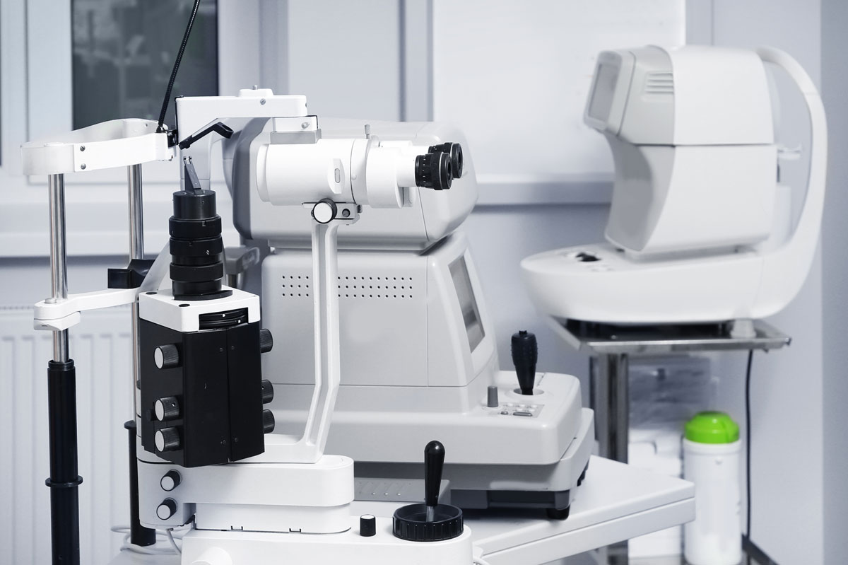
Humphrey® Field Analyzer – HFA II-i Series

Validated by more than 30 years of research, design and clinical experience, the Humphrey® Field Analyzer (HFA™) is the accepted standard of care for the diagnosis and management of neurological diseases, including glaucoma. The Humphrey Field Analyzer Guided Progression Analysis™ (GPA™) software differentiates statistically significant progression of visual field loss from random variability. The analysis is based upon detailed empirical knowledge of the variability found at various stages of glaucomatous visual field loss through information acquired in extensive multi-center clinical trials worldwide.

Icare Tonometer

Our vision is to provide patients with a comfortable, enjoyable experience while collecting the very best data to ensure long-term health. The Icare Tonometer allows us to test a patient’s intraocular pressure (IOP) to screen for glaucoma without needing to use an eye drop or that dreaded puff of air.

OCT with Angiovue for Glaucoma

The OCT is the best management tool available for glaucoma. Today’s OCT glaucoma applications include RNFL, ONH and angle assessment as well as the new Ganglion Cell Analysis (GCA). With angle imaging and central corneal thickness measurements, you have the world’s standard-setting comprehensive tool for glaucoma structural assessment. Guided Progression Analysis™ (GPA) provides a powerful tool for determining change for RNFL and optic nerve head parameters over time. Glaucoma is a condition that can lead to blindness if left undetected. Patients are without symptoms until the final stages so early detection is key.

OCT with Angiovue for Retinal Disease

This state-of-the-art non-invasive diagnostic instrument allows us to view cross sections of the retina, unobtainable with the naked eye. The OCT provides the framework to comprehensively assess a patient’s retinal condition and monitor progression or change in the future, prior to any symptoms being present in the patient. Three-dimensional views can be used for in- depth analysis, treatment planning and patient education of various conditions: macular degeneration, diabetic retinopathy, central serous, choroidal nevi and retinal detachments to name a few. Studies show that it can even be an early detector for multiple sclerosis.

The Angiovue feature provides the ability to quickly visualize the presence or absence of retinal vessels and assess new information about the microvasculature with extraordinary detail. This information helps form a complete picture of a patient’s disease state and their treatment options with conditions such as macular degeneration or diabetic retinopathy.

OPD 3D Wave

The OPD 3D Wave combines advanced technologies into one easy-to-use refractive and corneal diagnostic workstation. Now, with a single device, we can obtain day and night refraction, corneal topography, optical path difference (OPD), wave front analysis and early detection of corneal diseases such as keratoconus. Access to multiple data points results in a more accurate, custom refraction in less time and without the laborious process of innumerable options of “which is better, one or two?” You will see better than ever before and enjoy the process of getting there.

Optomap

The Optomap gives us a 200-degree, 3-D wrap of the back of your eye, allowing us to detect 40% more retinal conditions than dilation alone. It is all about access to information. The autoflourescence allows us to see photoreceptor damage often before structural changes present. The structural image uses red and green light versus white light, which can hinder the ability to detect conditions. The use of red and green light allows us to separate out the layers of the retina, looking at pigmentary changes such as a freckle or retinal splitting with retinoschisis. A patient benefit is that dilation drops are not required to get the images. The Optomap exam is fast, painless and comfortable. Nothing touches your eye at any time and it is suitable for the whole family. Images are available immediately for review. You can see your retina—exactly what Dr. Lambright sees—even in a 3-D animation. Early detection means successful treatments can be administered and reduces the risk to your sight and health.

TRS-5100 Marco Automated Refracting System

Our patients have access to the TRS-5100 automated refracting system, replacing the traditional manual refractor. This system enhances the patient experience through a faster, more modern exam. The TRS-5100 automated refracting system generates extremely accurate and reliable refractions, in a smoother, more patient friendly sequence, and allows the patient to experience prescription changes with the click of a button, while smoothly integrating with your electronic medical record.